|
|
|
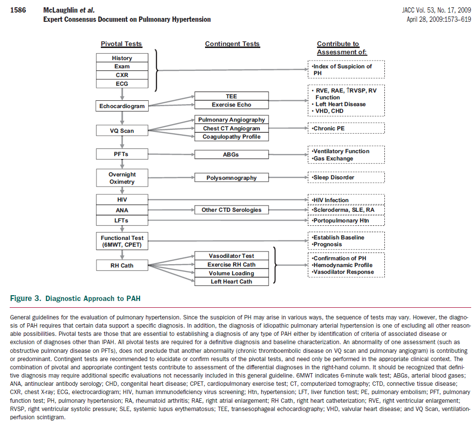
Pulmonary Arterial Hypertension: Tables and Figures from the 2009 ACC/AHA/ACCP Expert Consensus Document |
|
|
|
|
|
|
|
Postero-anterior and lateral chest X-ray (top) shows decreased peripheral lung vascular markings, hilar pulmonary artery prominence, and right ventricular enlargement of a patient with idiopathic PAH. ECG (bottom) of the same patient reveals right atrial enlargement, right ventricular hypertrophy and strain, and right axis deviation of the QRS complex. ECG indicates electrocardiogram; and PAH, pulmonary arterial hypertension. |
|
|
|
|
|
|
|
|
|
|